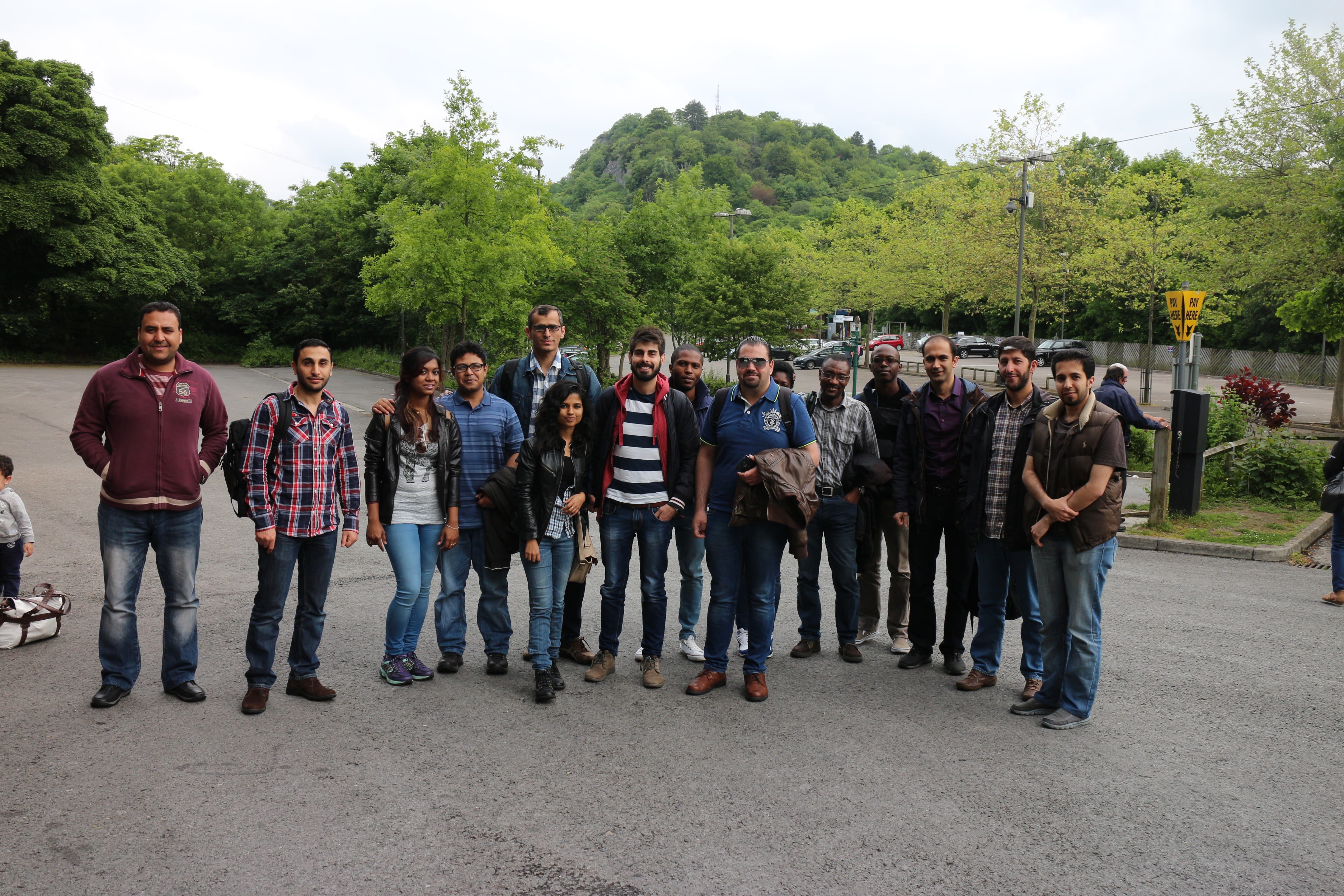
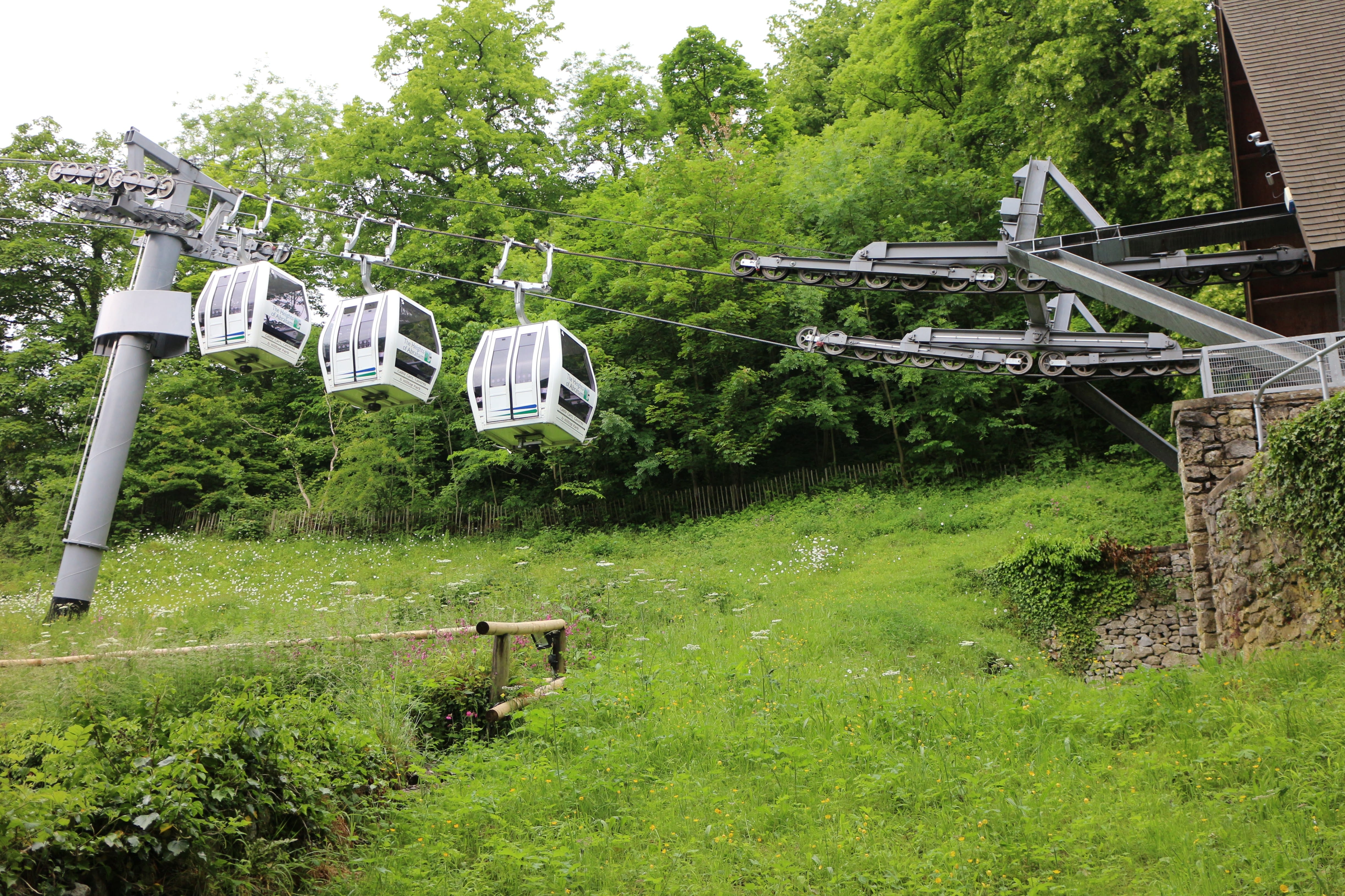
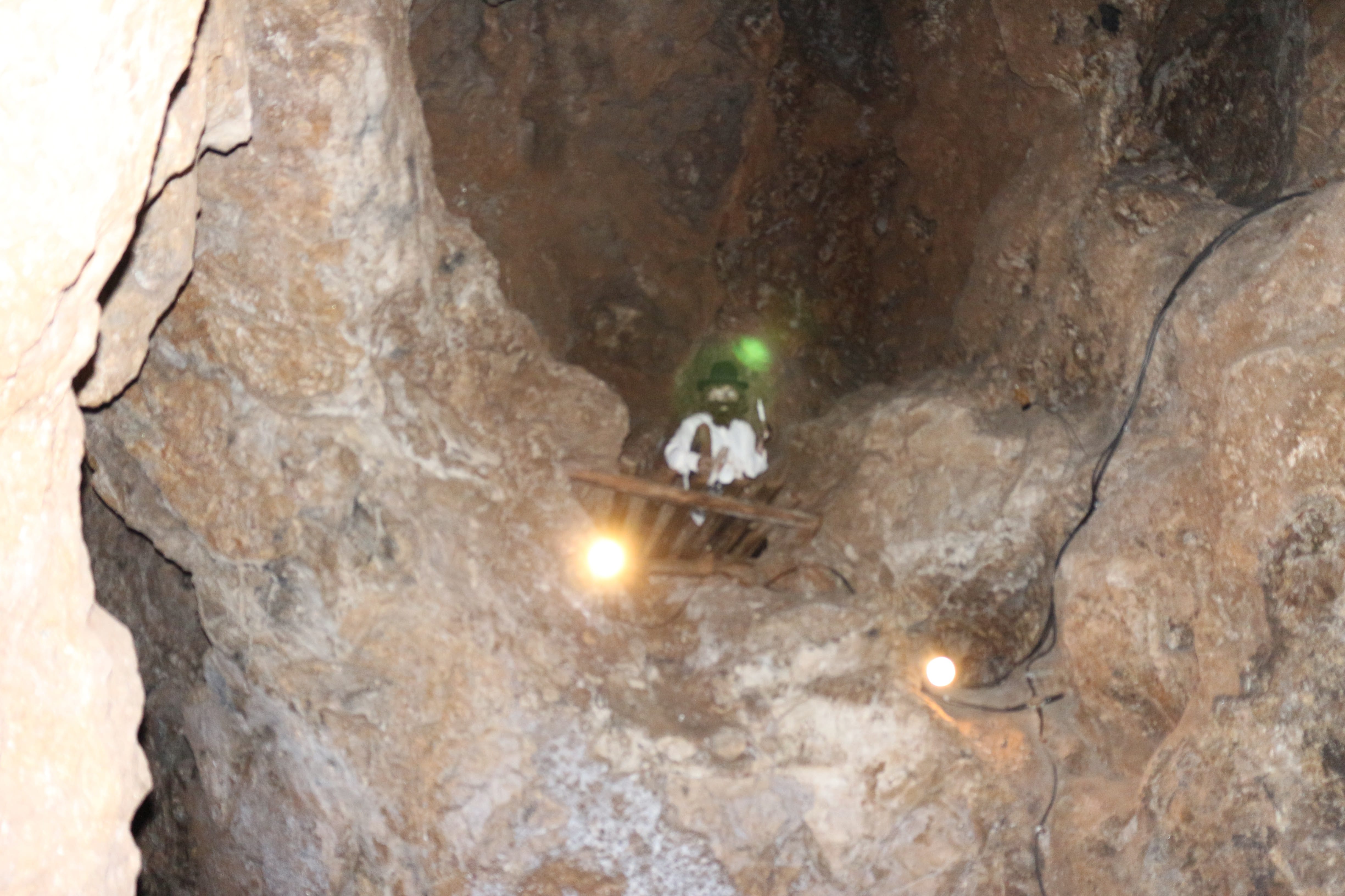
Another interesting day trip to Abraham Heights, Derby. 14th June 2015.
All got the cable cars, enjoyed tours inside old mining sites, and fantastic views from the top.
School of computer Science
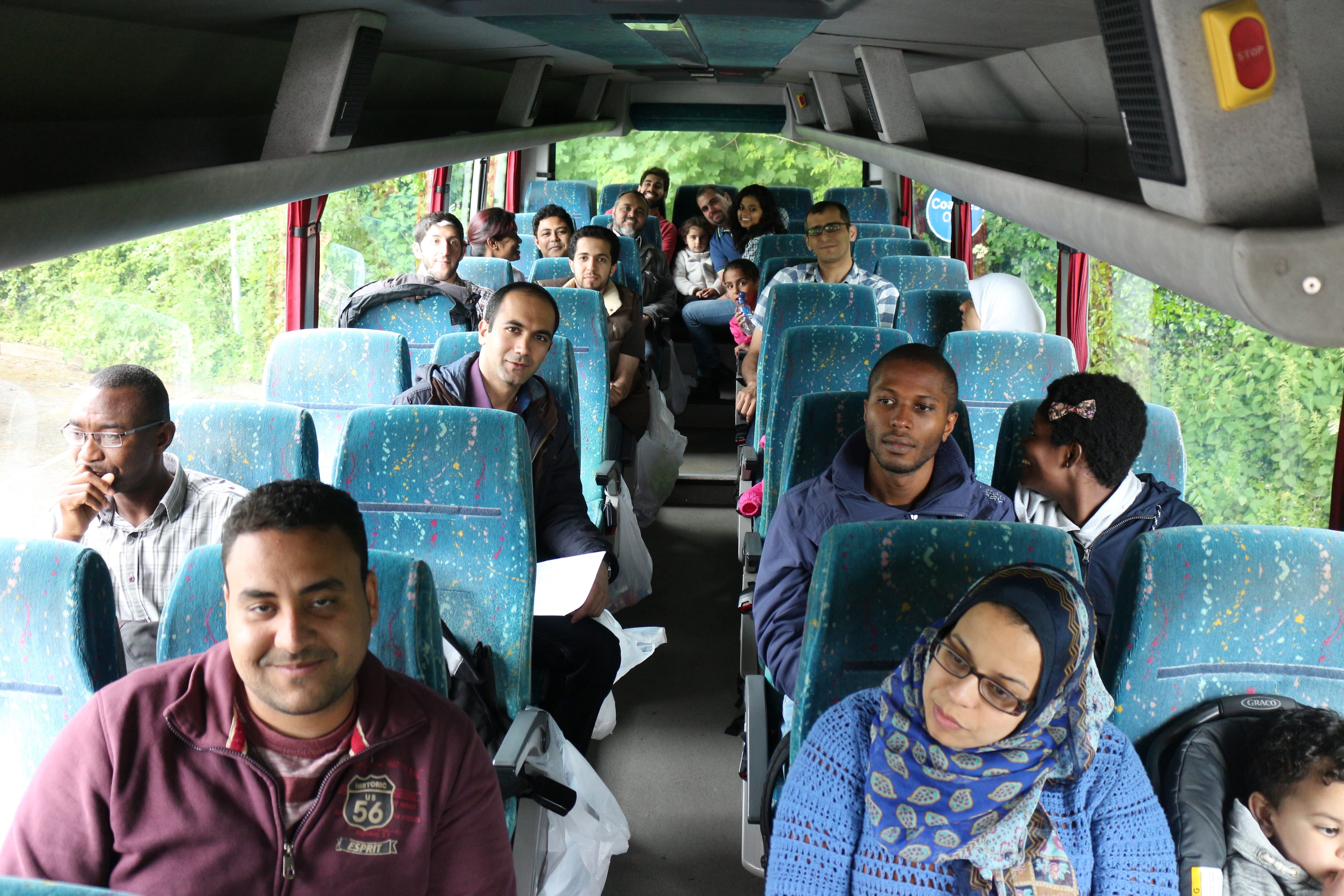
Another interesting day trip to Abraham Heights, Derby. 14th June 2015.
All got the cable cars, enjoyed tours inside old mining sites, and fantastic views from the top.
The monthly PGRs Research Presentations was held on Wed. 10th June, 2pm, Room MC3108.
This session we had the following presentations:
| Title: “Effects of Environmental Changes on Aggregation with Robot Swarm“. | Title: “Computer-aided Liver lesion detection and classification” | |
|
By: Farshad Arvin |
By: Hussein Alahmer |
|
| Abstract: Aggregation is one of the most fundamental behaviors that has been studied in swarm robotic researches for more than two decades. The studies in biology revealed that environment is a very important factor especially in cue-based aggregation in which a heterogeneity in the environment such as a heat or light source act as a cue indicating an optimal aggregation zone. In swarm robotics, studies on cue-based aggregation mainly focused on different methods of aggregation and different parameters such as population size. Although of utmost importance, effects of environmental factors have not been studied extensively. In this work, we study the effect of different environmental factors such as size and texture of aggregation cues and speed of the changes on a dynamic environment using real robots. We used aggregation time and size of the aggregate as the two metrics and evaluated the performance of the swarm aggregation in static and dynamic environments. The results of the performed experiments illustrate how environmental changes influence the performance of a swarm aggregation. |
Abstract: Liver cancer is one of the major death factors in the world. Transplantation and tumor resection are two main therapies in common clinical practice. Both tasks need image assisted planning and quantitative evaluations. An efficient and effective automatic liver segmentation is required for corresponding quantitative evaluations. Computed Tomography (CT) is highly accurate for liver cancer diagnosis. Manual identification of hepatic lesions done by trained physicians is a time-consuming task and can be subjective depending on the skill, expertise and experience of the physician. Computer aided classification of liver tumors from abdominal Computer Tomography (CT) images requires segmentation and analysis of tumor. Automatic segmentation of tumor from CT images is difficult, due to the size, shape, position and presence of other objects with the same intensity present in the image. The proposed system automatically segment liver from abdominal CT and detect hepatic lesions, then classifies the lesion into Benign or Malignant. The method uses Fuzzy C Means (FCM) clustering and region growing technique. The effectiveness of the algorithm is evaluated by comparing automatic segmentation results to the manual segmentation results. Quantitative comparison shows a close correlation between the automatic and manual as well as high spatial overlap between the regions-of-interest (ROIs) generated by expert radiologist and proposed system. |
|
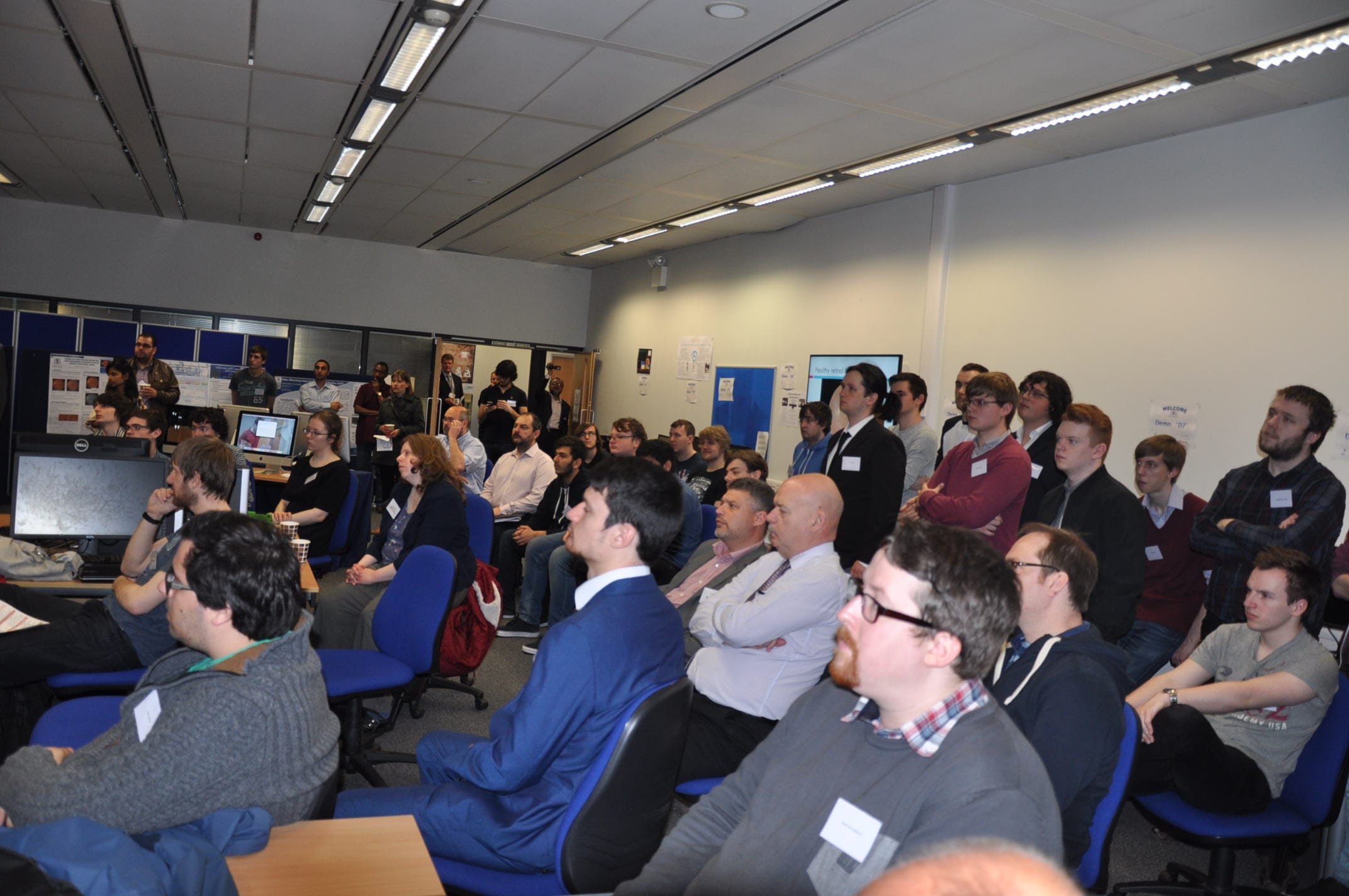
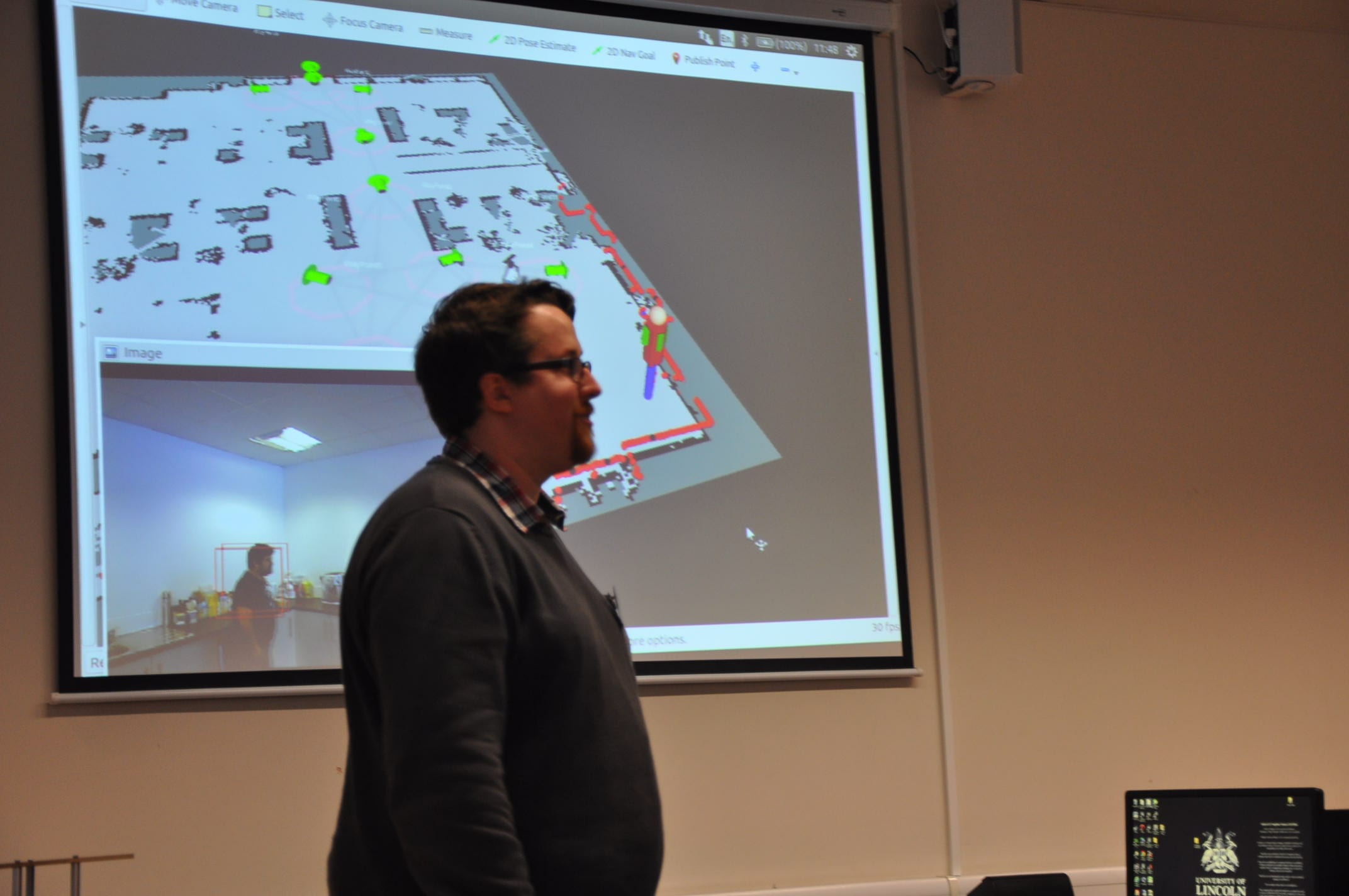
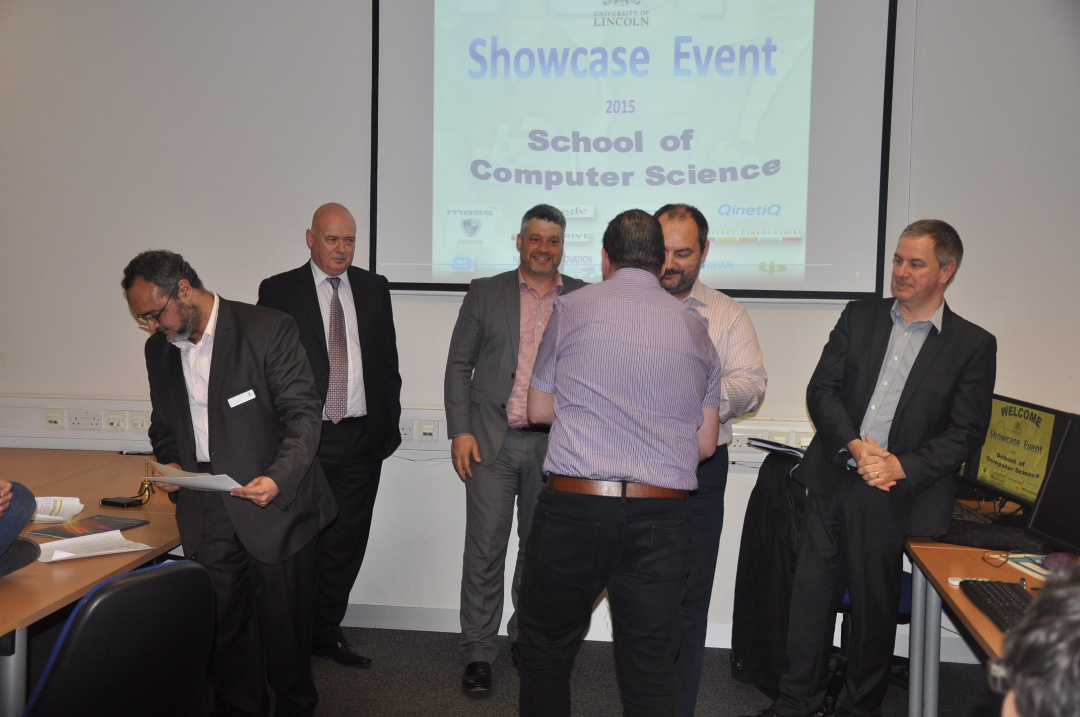
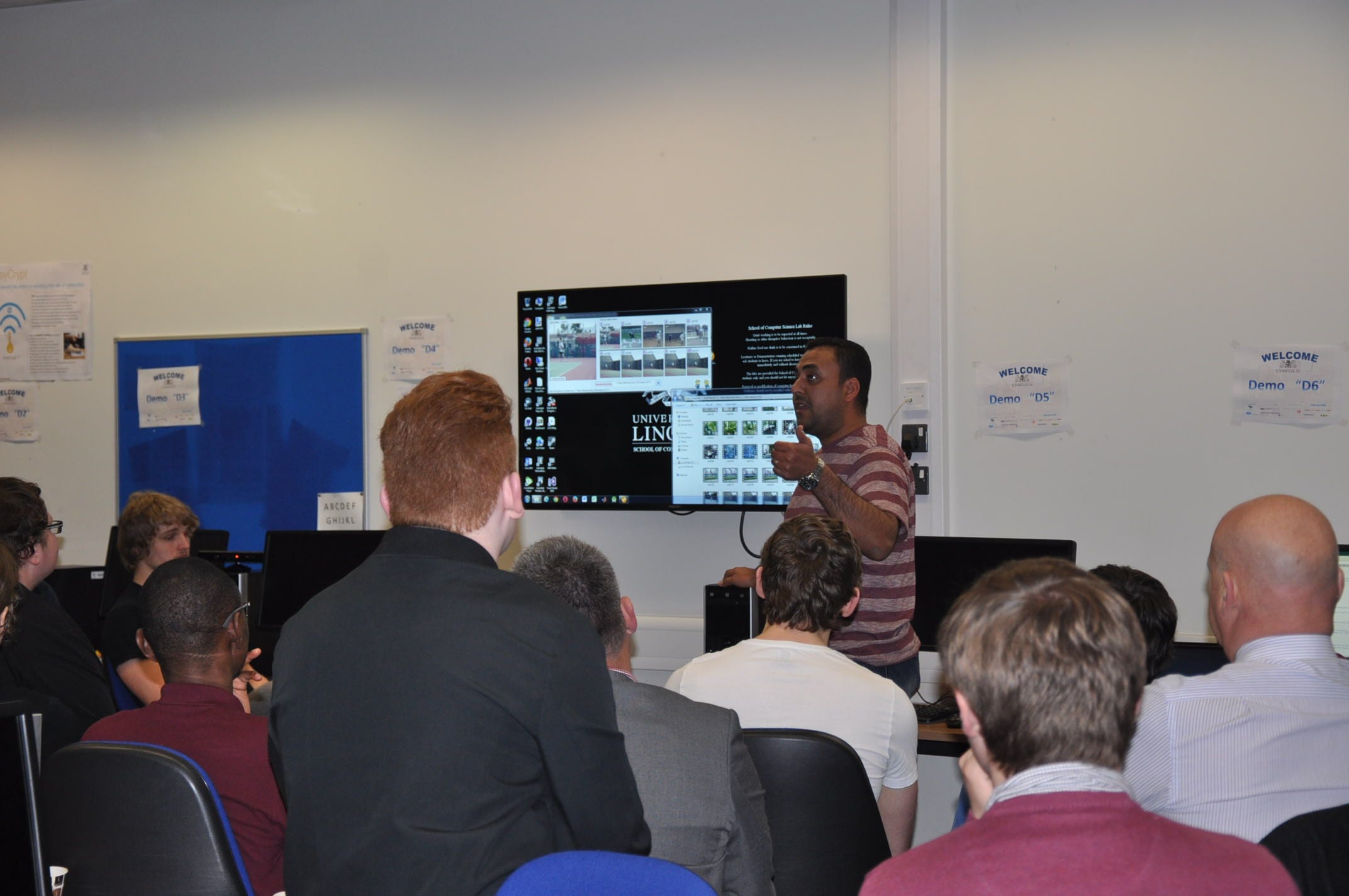
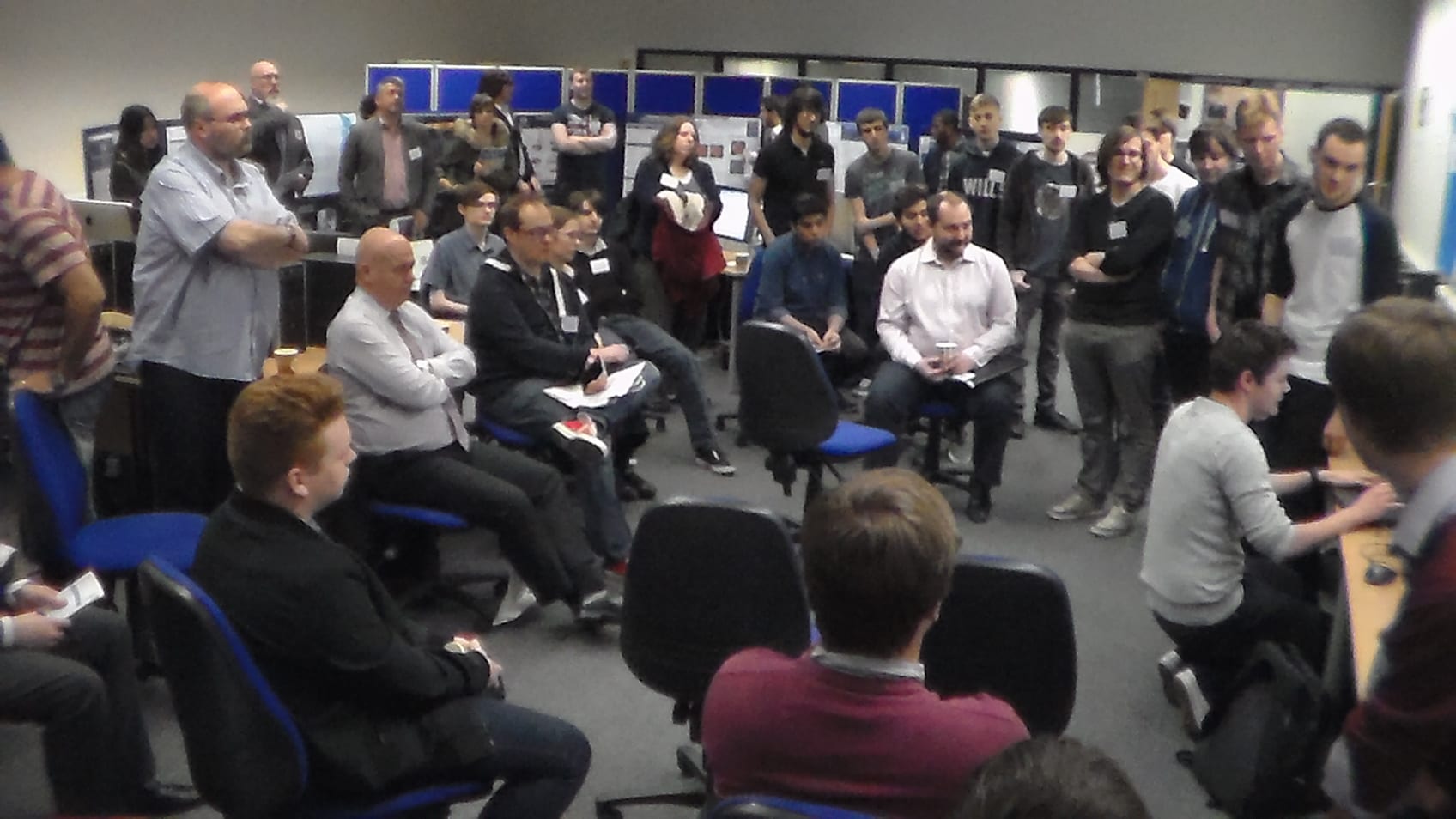
Postgraduates by Research (PGRs) presented and showed their research work in the Annual Showcase Event for the School of Computer Science, University of Lincoln. (6th May). The event spread over all the day with visitors and companies representatives. Guests praised the high quality of presented work and demos. A high level of engagement and discussions took place during the day.
The panel had difficulty deciding on the winners. But finally agreed on:
* Christian Dondrup.
* Saddam Bekhet.
* Alaa Al-Zoubi
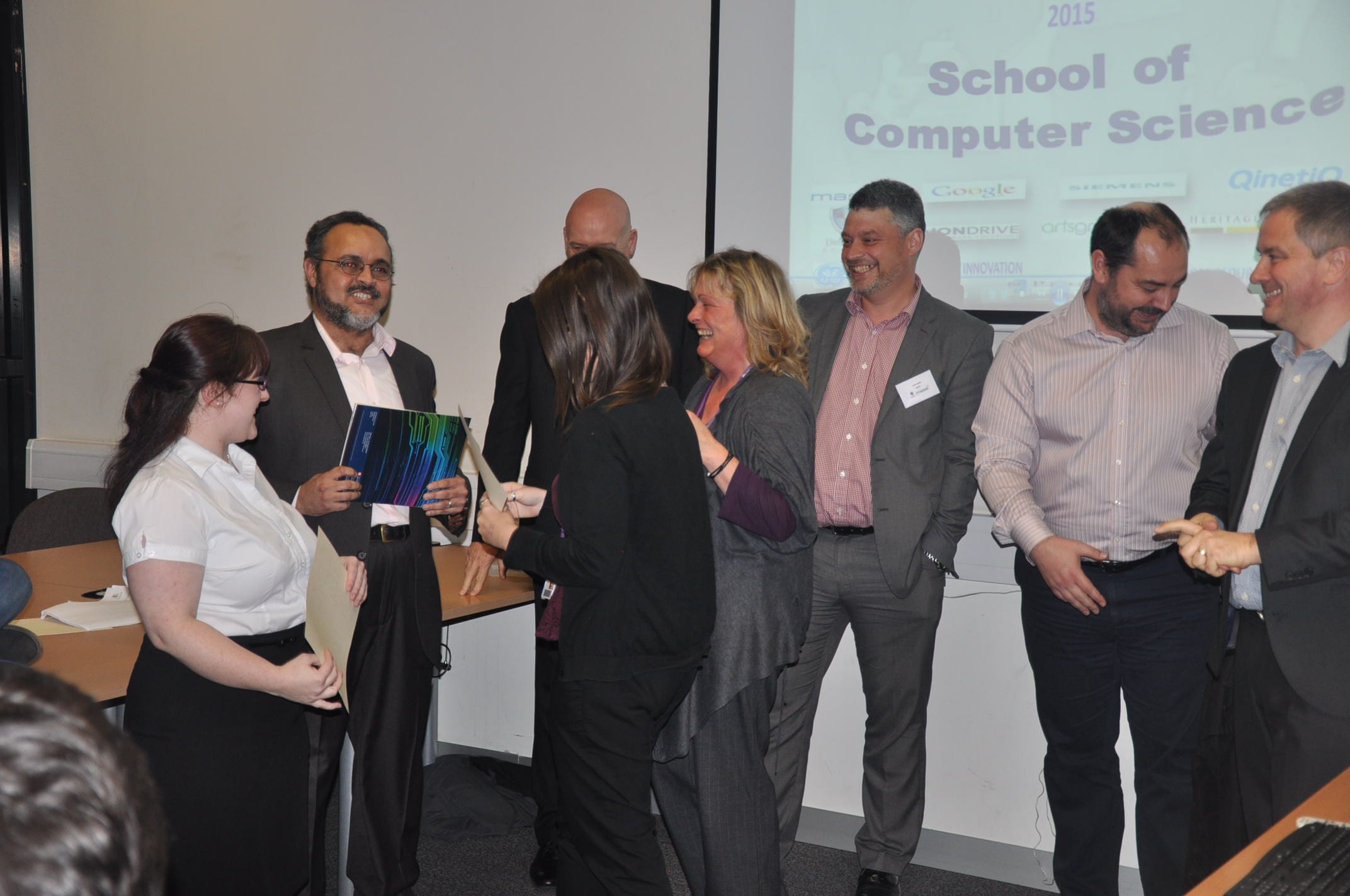
Thank to all participants, contributors, and all who helped in the event (and in the preparation).
The monthly PGRs Research Presentations was held on Wed. 11th March, 2pm, Room MC3108.
This session we had the following presentations:
| Title: “Retinal Vascular Measurement“. | Title: “4D Lifelong Exploration of Dynamic Environments” | |
|
By: Francesco Caliva |
By: Joao Santos |
|
| Abstract: Several studies have shown that systemic diseases affect blood vessels’ geometry. Retina is a window in the vascular system, thus fundus images can be adopted to diagnose or evaluate pathological conditions. Segmentation algorithms are not able to completely segment blood vessels. This failure results in a set of disconnected vascular segments. Reconstructing the whole network has crucial importance. At this aim, in this work, implicit neural cost functions have been adopted to evaluate how the segments can be joined. In this talk I will present my current and future work. | Abstract: We present a novel 4D lifelong exploration method for dynamic, human populated environments. In contrast to other exploration methods that model the environment as being static, our spatio-temporal exploration method creates and maintains a world model that not only represents the environment’s 3D structure, but also its dynamics over time.The predictive ability of the 4D spatio-temporal model allows the exploration method not only where, but also when to make environment observations. To validate our method, a mobile robot was deployed over 5 days in an office environment, and the proposed method was compared against a static approach. The results show that through understanding of the environment dynamics, the spatio-temporal exploration algorithm could predict which locations were going to change at a specific time and used this knowledge to guide the robot. This allowed our spatio-temporal exploration method to gather more information that the exploration method that relied on a static environment model. |
|
The monthly PGRs Research Presentations was held on Wed. 11th February, 2pm, Room MC3108.
This session we had the following presentations:
| Title: “Designing, Developing and Evaluating Mobile and Social Technology to Improve Sleep Quality“. | Title: “Brain Tumour Grading in Different MRI Protocols using SVM on Statistical Features ” | |
|
By: Ibraheem Alnejaidi |
By: Mohammadreza Soltaninejad |
|
| Abstract: | Abstract: Brain tumours are caused by abnormal and uncontrolled growing of the cells inside the brain or spinal canal. Regarding to the World Health Organization (WHO) grading system, the tumours are graded from I to IV, corresponding to least advanced to the most advanced diseases, respectively. In our work a feasibility study of brain MRI dataset classification, using ROIs which have been segmented either manually or through a superpixel based method in conjunction with statistical pattern recognition methods is performed. The aim is classifying tumour grades II, III and IV using different MRI acquisition protocols i.e. FLAIR, and T2. We found by using the Leave-One-Out method that the combination of the features from the 1st and 2nd order statistics, achieved high classification accuracy in pair-wise grading comparisons. | |
|